Zeiss Cirrus HD-OCT 4000

General details
Carl Zeiss Cirrus 4000 HD-OCT
YOM: 2014
Condition: Excellent
Installed software: software of Model 500/5000 Version 10.0.1
To pictures:
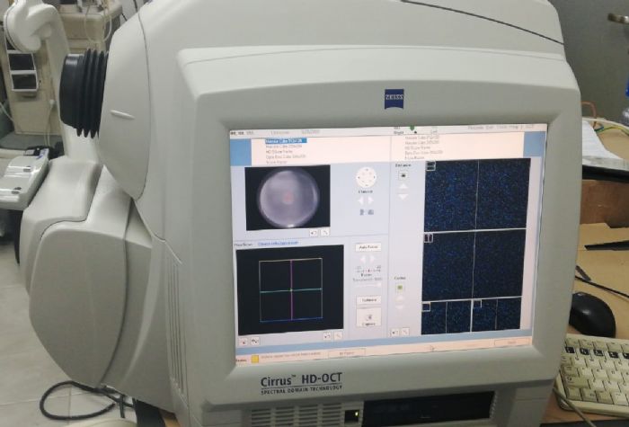
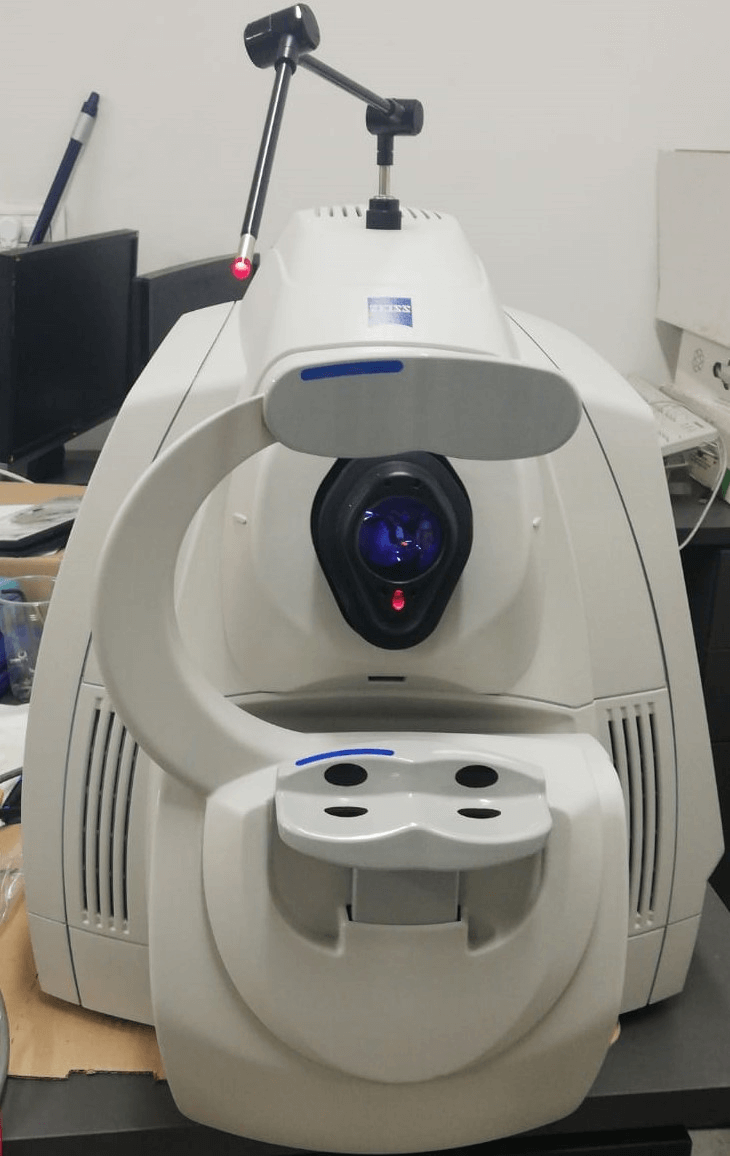
The ZEISS Cirrus HD-OCT 4000 (Cirrus HD-OCT or Cirrus) enable examination of the posterior and anterior of the eye at an extremely fine spatial scale, without surgical biopsy or even any contact with the eye. The Cirrus HD-OCT builds on and refines the retinal imaging technology first introduced with the ZEISS Stratus OCT™. HD-OCT stands for "high-definition optical coherence tomography." Employing the advanced imaging technology of spectral domain optical coherence tomography, Cirrus HD-OCT acquires OCT data about 70 times faster (27,000 vs. 400 A-scans per second) and with better resolution (5 μm vs. ~10 μm axial resolution in tissue), compared to first-generation OCT technology. Cirrus acquires whole cubes of OCT image data, composed of hundreds of line scans, in about the same time as Stratus acquires a six-line scan. You can view these data cubes in three planes, or through three dimensions, giving you access to an extensive amount of retinal image data in one scan. Intended UseThe Cirrus HD-OCT with Retinal Nerve Fiber Layer (RNFL), Macular, Optic Nerve Head, and Ganglion Cell Normative Databases is indicated for in-vivo viewing, axial cross-sectional, and three-dimensional imaging and measurement of anterior and posterior ocular structures. Indications for UseThe Cirrus HD-OCT is a non-contact, high resolution tomographic and biomicroscopic imaging device. It is indicated for in-vivo viewing, axial cross-sectional, and three-dimensional imaging and measurement of anterior and posterior ocular structures, including cornea, retina, retinal nerve fiber layer, ganglion cell plus inner plexiform layer, macula, and optic nerve head. The Cirrus normative databases are quantitative tools for the comparison of retinal nerve fiber layer thickness, macular thickness, ganglion cell plus inner plexiform layer thickness, and optic nerve head measurements to a database of normal subjects. The Cirrus HD-OCT is intended for use as a diagnostic device to aid in the detection and management of ocular diseases including, but not limited to, macular holes, cystoid macular edema, diabetic retinopathy, age-related macular degeneration, and glaucoma. Note: The Cirrus HD-OCT is not intended to be used as the sole diagnostic for disease. Essential PerformanceThe Essential Performance of the instrument is to provide accurate measurements of anterior and posterior ocular structure. Patient PopulationThe Cirrus HD-OCT may be used on all adults in need of diagnostic evaluation of the eye. This includes (but is not limited to) patients with the following disabilities or challenges: • Wheelchair user• Very low or not measurable visual acuity• Fixation problems• Postural problems• Deafness• Large body, but not those above 99th percentile based on anthropomorphic data There is a general requirement that the patient be able to sit upright and be able to place their face in the chin and forehead rest of the instrument (with or without supplemental human or mechanical support).Cirrus HD-OCT is designed for in-vivo viewing, axial cross-sectional, and three-dimensional imaging and measurement of anterior and posterior ocular structures. Specifications: HD-OCT ImagingMethodology: Spectral Domain OCTOptical Source: superluminescent diode (SLD), 840 nmOptical Power: < 725 μW at the corneaScan Speed: 27,000 A-scans per secondA-Scan depth: 2.0 mm (in tissue), 1024 pointsAxial resolution: 5 μm (in tissue)Transverse resolution: 15 μm (in tissue) Fundus ImagingMethodology: Line scanning ophthalmoscopeLive Fundus Image: During alignment and during OCT scanOptical Source: Superluminescent diode (SLD), 750 nmOptical Power: < 1.5 mW at the corneaField of View: 36 degrees W x 30 degrees HFrame rate: >20 HzTransverse resolution: 25 μm (in tissue) Iris ImagingMethodology: CCD CameraResolution: 1280 x 1024Live iris image: During alignment Electrical, Physical and EnvironementalWeight: 38kg (83 lbs)Dimensions: 65L x 44W x 53H (cm)Fixation: Internal, externalInternal fixation focus adjustment: -20D to +20D (diopters)Input devices: Keyboard, mouseElectrical rating (115V) Single Phase, 100/120V~ systems:50-60Hz, 5AFuse rating (115V) T 5A 250VElectrical rating (230V) Single Phase, 220/240V~ systems:50-60 Hz, 2.5AFuse rating (230V) Fuse rating: T 5A 250VConvenience Receptacle output ratings 115V~, 0.5 A Max, 50-60 HzTemperature (transport and storage) -40º to +70º CRelative humidity (transport and storage) 10% to 100%, including condensationAtmospheric pressure (transport and storage): 500 hPa to 1060 hPaTemperature (operation) +10º to +35º CRelative humidity (operation) 30% to 75%, excluding condensationAtmospheric Pressure (operation) 700 hPa to 1060 hPa Computer • High performance multi-core processor• Internal storage: > 80,000 scans• CD-RW, DVD-ROM drive• Integrated 15" color flat panel display
Pictures:

YOM: 2014
Condition: Excellent
Installed software: software of Model 500/5000 Version 10.0.1
To pictures:
The ZEISS Cirrus HD-OCT 4000 (Cirrus HD-OCT or Cirrus) enable examination of the posterior and anterior of the eye at an extremely fine spatial scale, without surgical biopsy or even any contact with the eye. The Cirrus HD-OCT builds on and refines the retinal imaging technology first introduced with the ZEISS Stratus OCT™. HD-OCT stands for "high-definition optical coherence tomography." Employing the advanced imaging technology of spectral domain optical coherence tomography, Cirrus HD-OCT acquires OCT data about 70 times faster (27,000 vs. 400 A-scans per second) and with better resolution (5 μm vs. ~10 μm axial resolution in tissue), compared to first-generation OCT technology. Cirrus acquires whole cubes of OCT image data, composed of hundreds of line scans, in about the same time as Stratus acquires a six-line scan. You can view these data cubes in three planes, or through three dimensions, giving you access to an extensive amount of retinal image data in one scan. Intended UseThe Cirrus HD-OCT with Retinal Nerve Fiber Layer (RNFL), Macular, Optic Nerve Head, and Ganglion Cell Normative Databases is indicated for in-vivo viewing, axial cross-sectional, and three-dimensional imaging and measurement of anterior and posterior ocular structures. Indications for UseThe Cirrus HD-OCT is a non-contact, high resolution tomographic and biomicroscopic imaging device. It is indicated for in-vivo viewing, axial cross-sectional, and three-dimensional imaging and measurement of anterior and posterior ocular structures, including cornea, retina, retinal nerve fiber layer, ganglion cell plus inner plexiform layer, macula, and optic nerve head. The Cirrus normative databases are quantitative tools for the comparison of retinal nerve fiber layer thickness, macular thickness, ganglion cell plus inner plexiform layer thickness, and optic nerve head measurements to a database of normal subjects. The Cirrus HD-OCT is intended for use as a diagnostic device to aid in the detection and management of ocular diseases including, but not limited to, macular holes, cystoid macular edema, diabetic retinopathy, age-related macular degeneration, and glaucoma. Note: The Cirrus HD-OCT is not intended to be used as the sole diagnostic for disease. Essential PerformanceThe Essential Performance of the instrument is to provide accurate measurements of anterior and posterior ocular structure. Patient PopulationThe Cirrus HD-OCT may be used on all adults in need of diagnostic evaluation of the eye. This includes (but is not limited to) patients with the following disabilities or challenges: • Wheelchair user• Very low or not measurable visual acuity• Fixation problems• Postural problems• Deafness• Large body, but not those above 99th percentile based on anthropomorphic data There is a general requirement that the patient be able to sit upright and be able to place their face in the chin and forehead rest of the instrument (with or without supplemental human or mechanical support).Cirrus HD-OCT is designed for in-vivo viewing, axial cross-sectional, and three-dimensional imaging and measurement of anterior and posterior ocular structures. Specifications: HD-OCT ImagingMethodology: Spectral Domain OCTOptical Source: superluminescent diode (SLD), 840 nmOptical Power: < 725 μW at the corneaScan Speed: 27,000 A-scans per secondA-Scan depth: 2.0 mm (in tissue), 1024 pointsAxial resolution: 5 μm (in tissue)Transverse resolution: 15 μm (in tissue) Fundus ImagingMethodology: Line scanning ophthalmoscopeLive Fundus Image: During alignment and during OCT scanOptical Source: Superluminescent diode (SLD), 750 nmOptical Power: < 1.5 mW at the corneaField of View: 36 degrees W x 30 degrees HFrame rate: >20 HzTransverse resolution: 25 μm (in tissue) Iris ImagingMethodology: CCD CameraResolution: 1280 x 1024Live iris image: During alignment Electrical, Physical and EnvironementalWeight: 38kg (83 lbs)Dimensions: 65L x 44W x 53H (cm)Fixation: Internal, externalInternal fixation focus adjustment: -20D to +20D (diopters)Input devices: Keyboard, mouseElectrical rating (115V) Single Phase, 100/120V~ systems:50-60Hz, 5AFuse rating (115V) T 5A 250VElectrical rating (230V) Single Phase, 220/240V~ systems:50-60 Hz, 2.5AFuse rating (230V) Fuse rating: T 5A 250VConvenience Receptacle output ratings 115V~, 0.5 A Max, 50-60 HzTemperature (transport and storage) -40º to +70º CRelative humidity (transport and storage) 10% to 100%, including condensationAtmospheric pressure (transport and storage): 500 hPa to 1060 hPaTemperature (operation) +10º to +35º CRelative humidity (operation) 30% to 75%, excluding condensationAtmospheric Pressure (operation) 700 hPa to 1060 hPa Computer • High performance multi-core processor• Internal storage: > 80,000 scans• CD-RW, DVD-ROM drive• Integrated 15" color flat panel display
Pictures: